

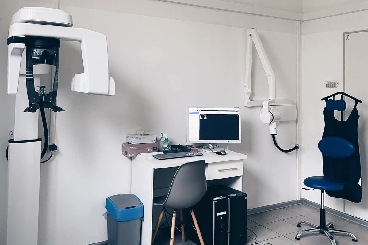
Precise diagnosis is the foundation of effective dental treatment. At Dobry Dentysta we use digital radiography — periapical X-rays, panoramic X-rays and CBCT 3D cone beam scans — for a detailed assessment of the teeth, jawbones, sinuses and temporomandibular joints.
Types of dental X-rays
Several types of X-ray are used in dental diagnostics, chosen according to the clinical situation. The simplest is a periapical (dental) X-ray, which shows a single tooth or a small area — the standard examination for suspected decay or during root canal treatment. A panoramic X-ray (OPG) covers the whole dental arch together with the branches of the lower jaw and the maxillary sinuses and is used mainly when planning orthodontic and surgical treatment. The most advanced method is CBCT 3D cone beam tomography, which gives a three-dimensional image of the jawbones — it is essential for implant planning, oral surgery and the diagnosis of more complex conditions.
When is a CBCT 3D scan necessary?
A CBCT 3D scan is performed in several specific clinical situations. It is essential when planning implants, as it allows precise assessment of bone volume and density and locates the mandibular nerve. It is also used to diagnose impacted wisdom teeth, especially those close to the inferior alveolar nerve, and in advanced root canal treatment, where it shows the number, course and curvature of the root canals precisely. CBCT is also used to assess bone lesions such as cysts or granulomas when planning surgery, and in orthodontics and orthognathic surgery, where it enables three-dimensional analysis of the bones and teeth.
X-ray safety
Digital X-ray equipment emits a radiation dose several times lower than analogue equipment. The dose from a dental X-ray is very small — lower than the dose received on a short flight. Pregnant women should tell the dentist — we take X-rays only when clearly indicated.
Digital image = better diagnosis
A digital X-ray sensor gives an immediate result without developing. The image can be enlarged and its contrast and brightness adjusted, making it much easier to diagnose early decay, root fractures or inflammation at the root tips.